UMHS /

    Anatomy Lab

    Anatomy Lab


    The University of Medicine and Health Sciences is proud to have the best Anatomy lab in the Caribbean. Even the largest medical schools in the United States would be envious of the anatomy lab and facilities we have! The gross anatomy lab and neuroanatomy laboratories are housed in our 11,000 sq. ft. anatomy facility, which is equipped with cutting-edge audio-visual technology. There are 20 dissection platforms in the gross anatomy lab where students have a 6 to 1 cadaver ratio. While in neuroanatomy the ratio is even lower at 4 to 1.

    For demonstration purposes, a prosected cadaver is used in class. A high-resolution camera records even the slightest details and presents them on the 55-inch LED monitors that are located at the end of every student's cadaver work table. This allows all students to see all the important dissection details while working on their cadavers.

    During anatomy lab sessions, students can use computer software at each cadaver table to obtain anatomical reference information. Students can also view pertinent information offered by the professor in a variety of formats while dissecting, including Powerpoint and ELMO® presentations. Power poles are present at each cadaver table enabling students who want to use their laptops or tablets to access our library of anatomical photos.


    Get a 360 degree look inside our Anatomy lab and other state-of-the-art facilities

    Take a virtual tour
    Anatomy lab
    ×

    Some quick points about Anatomy Labs

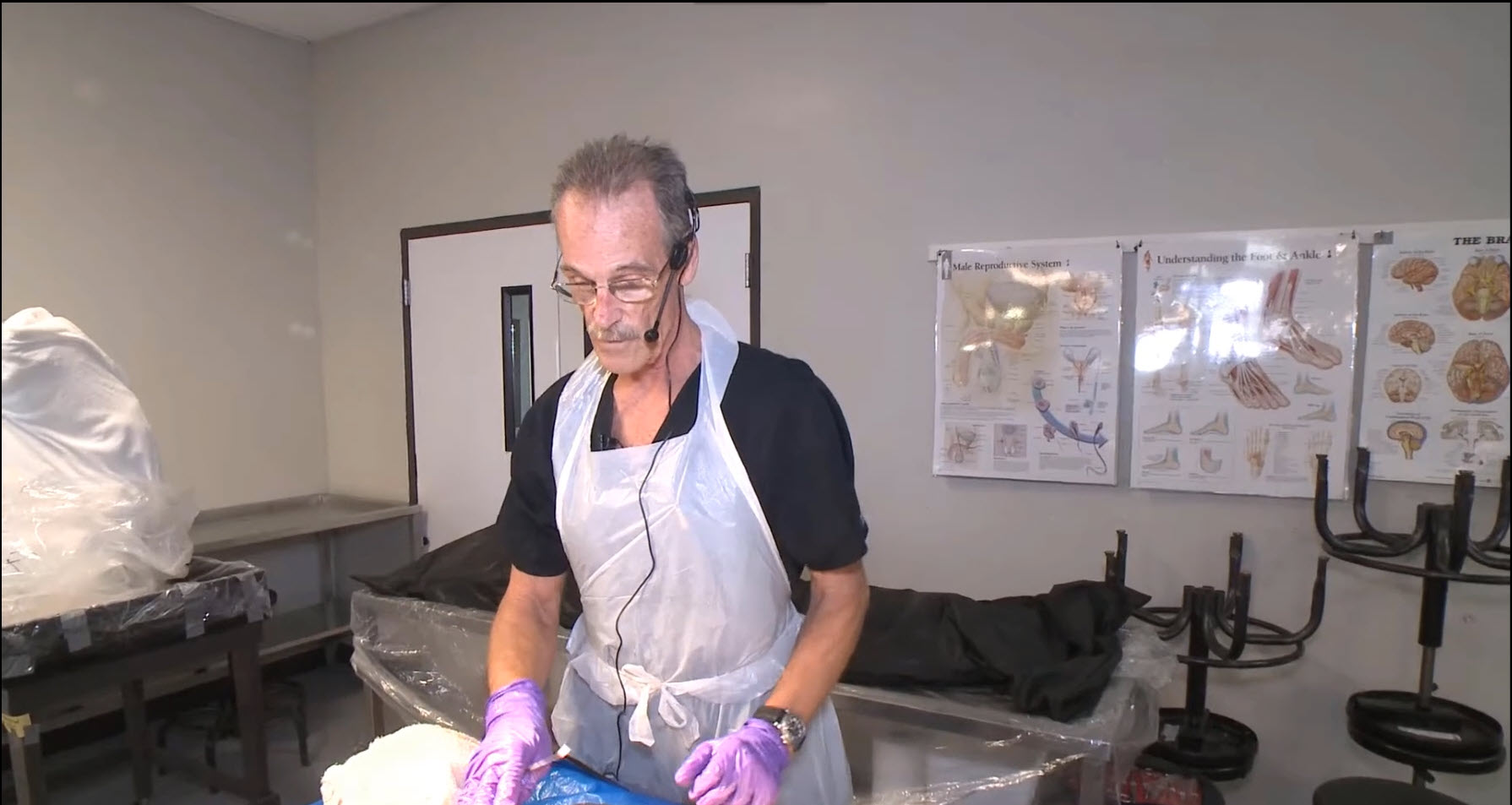
    Anatomy forms the foundation of medical knowledge. It's a fundamental, essential component of medical education that cannot be replaced. At The University of Medicine and Health Sciences, our students perform cadaver dissection. This is a very important step in a medical student's education as gaining an understanding of human variability is not replaceable by using video technology.

    State of Art Anatomy Lab facility

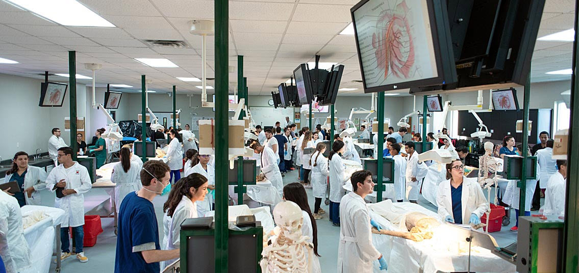
    State of the art Anatomy lab

    The image on the right shows that no expense was spared during the construction of this 11,000 square foot facility from the dissection tables to the ventilation system. Notice that the stations are not crowded and advanced audio-visual systems allow everyone to follow along.

    FAQs

    Here are answers to the most common questions we get asked. 

    Anatomy labs are classrooms where a student, usually in a health medical-related field, is taught human clinical anatomy. Human anatomy lab is an important course in school for medical, dental, nursing, and even undergraduate students where they research and learn the structures of the body. This includes all the anatomical organs, ligaments, bones, arteries, veins, and connective tissue. For any student in undergraduate school or university, images and models are commonly used to teach anatomy lab. However, in most medical schools, students learn anatomy by dissecting human cadavers in an anatomy lab. This study process in school may also be augmented by the use of digital media and virtual simulations. The Covid-19 pandemic has temporarily limited cadaver dissection in the anatomy lab so some schools may have courses taught online.

    For most medical schools in the United States, completing a course with a gross anatomy lab prior to attending medical school is not required for application or matriculation (although doing your own research on pre-requisite requirements prior to applying to a school is always encouraged). However, previous exposure to human anatomy with an anatomy lab during undergraduate college or university, or even during high school, can help to prepare a student for the rigorous anatomical course during school. Any format- in-person, virtual, or online anatomy education may help to prepare students for anatomy courses in medical school, undergraduate university, or even other higher education schools.

    In anatomy lab, medical, and other health professions students have the opportunity to closely examine muscles, tendons, organs, nerves, and blood vessels that are taught in lectures. Most medical schools have future MD students research and conduct their own dissection of the anatomy cadavers. This is a vital part of education, as it is extremely important to learn the anatomical interactions between different structures of the body. For example, knowledge and education on the location and interaction of tendons and ligaments in the ankle are necessary to understand and treat ankle sprains and tears. In addition to dissection of anatomic cadavers, an anatomy lab may use virtual and video technology. Some school anatomy laboratories may also teach:

    •  Interpretation of important imaging techniques- such as Xray, CT/CAT scan, MRI, Ultrasound, etc.
    • Interpretation of histological slides- microscopic sections of tissues

    Using dissection in an Anatomy lab with human cadavers in university or school may be controversial. Some medical programs feel that learning anatomy can be properly done with the use of only images and models or by use of an Anatomage. However, research and feedback from students indicate that they prefer learning using cadavers and receive a better education compared to using only images and models. Dissection during school enables a student to better understand and research the interactions between structures as well as the location and path of certain anatomical structures inside the body. Most medical students also find it very helpful to see how different each body is. Cadaver dissection at an anatomy lab demonstrates and teaches students about common anatomical variants that may affect patient care. Another important aspect of the use of cadavers for anatomy education is that cadaver dissection can serve as an important way to conduct research. In particular, research on the prevalence of certain anatomical variants can be conducted on the cadavers used for anatomy lab education. The prevalence and location of these variations need to be known, particularly for surgeons that may encounter these variations during surgery.

    The smell inside a human anatomy lab in school is due to a preserving agent called Formaldehyde. Formaldehyde does not have a foul odor of decomposition; rather, it has a unique odor that lingers and may stick to your nostrils after you leave the school lab. Some individuals may also find that the formaldehyde may burn their eyes or nostrils while they are in close proximity to the agent. Additionally, the smell has been noted to 'linger' on clothes and hair after exiting the anatomy lab. (After exiting the gross anatomy lab, showering and sniffing potent scents such as coffee grounds seems to be a fantastic way to get rid of the lingering smell.)

    No. Taking a class with an Anatomy lab is not one of the most important classes for a student to take in preparation for taking the Medical College Admission Test (MCAT). Taking courses like Genetics, Cell Biology and Biochemistry will be more helpful.